Photonic crystal slabs (PhCS), i.e., two-dimensional (2D) photonic crystals with fifinite vertical dimension, have attracted much attention because of their potential applications to various optoelectronic devices and circuits. Defect cavities in these photonic devices can have high quality factor and small modal volume. The in-plane confifinement is achieved via Bragg scattering, while the index guiding prevents light leakage in the perpendicular direction. Low-threshold lasing has been realized in PhCS made of III-V semiconductors. They operate in the near infrared frequencies. Ultraviolet (UV) photonic crystal laser, however, has not been realized yet. This is because shorter wavelength requires smaller feature size, which is technologically challenging for commonly used wide band gap materials (e.g., GaN, ZnO). On the other hand, the demand for blue and UV compact laser sources has prompted enormous research effffort into wide band gap semiconductors. Compared with other wide band gap materials, ZnO has the advantage of large exciton binding energy (∼60 meV), that allows effiffifficient excitonic emission even at room temperature.
In this letter, we realized, for the fifirst time, ZnO photonic crystal lasers operating in the near-UV frequency at room temperature. We developed a procedure to fabricate 2D periodic structures in ZnO fifilms with the focused ion beam (FIB) etching. Post thermal annealing is employed to remove structural damage induced by FIB etching. Lasing is achieved in the strongly localized defect modes near the edges of photonic band gap by optical pumping. To explain the experimental results, we calculated the band structure of our samples using the 3D plane wave expansion method.
The samples are optically pumped by the third harmonics of a mode-locked Nd:YAG laser (355 nm, 10 Hz, 20 ps) at room temperature. A schematic sketch of the experimental setup is shown in Fig. 2. A 10× microscope objective lens (N.A.=0.25) is used to focus the pump beam to a 4 µm spot on one pattern, and also collect the emission from the pattern. Then the emitted light is focused by another lens into a UV fifiber, which is connected to a spectrometer with 0.13 nm spectral resolution. Since the sapphire substrate is double-side polished and transparent in both visible and UV frequencies, a 20× microscope objective lens (N.A.=0.40) is placed at the back side of the sample for simultaneous measurement of the spatial distribution of emission intensity. The pump light is blocked by a bandpass fifilter, while the image of lasing mode profifile is projected by the objective lens onto a UV sensitive CCD camera. The sample is also illuminated by a white light source so that we can identify the position of the lasing modes in the photonic lattice.
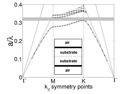
To understand our experimental results, we calculated the photonic band structures using the three-dimensional (3D) plane wave expansion method. Our ZnO PhCS is vertically asymmetric: with air on top and sapphire substrate at bottom. To apply the plane wave expansion method for photonic band calculation, we considered a super unit cell that consists of air, ZnO photonic crystal slab, sapphire substrate, ZnO photonic crystal slab and air (see the inset of Fig. 4). The guided modes con- fifined to the PhCS should lie inside the ZnO/substrate light cone and the air/ZnO light cone. If the substrate is thick enough, the guided modes in the two ZnO layers are decoupled, the symmetric and antisymmetric (with respect to the cell center plane – dashed line in the inset of Fig. 4) modes should be degenerate in eigenfrequency. This artifificial degeneracy is used as the check of consistency in our band structure calculations. In a symmetric PhCS (i.e. the substrate is replaced by air), the modes can be grouped into two classes: modes that have zcomponent of the magnetic fifield or electric fifield symmetric with respect to the center plane of the photonic layer. The complete stop bands for the guided modes of each class can exist independently. Low lying modes of the fifirst/second class are mainly transverse electric/magnetic (TE/TM) polarized. The presence of the substrate removes the symmetry. For our ZnO samples, we calculated the fifield components of the fifirst fifive bands with and without the sapphire substrate and found close resemblance in the spatial profifiles of the corresponding modes in the two cases. This is due to strong vertical confifinement of the guided modes. Therefore, the modes in the ZnO PhCS with the sapphire substrate can still be classifified as predominantly either TE-like or TM-like. It is known that the polarization of ZnO exciton emission is mostly perpendicular to the c-axis. Since the c-axis of our ZnO fifilm is normal to the fifilm/substrate interface, the emitted photons are coupled mainly into TE-like modes, as we confifirmed experimentally from the measurement of polarization of photoluminescence from the side of an unpatterned ZnO fifilm. Therefore, we can disregard the TM-like bands in the band structure calculation. For an appropriate choice of lattice parameters, a complete photonic band gap exists between the fifirst two TE-like bands. Fig. 4 shows an example of the calculated photonic band structures for a =130 nm and r/a = 0.25. For this system there exist a photonic band gapfrom 396 nm to 415 nm. For PhCS with a =115 nm and r/a = 0.25, a narrower gap exists between 363 nm and 372 nm. At room temperature, the ZnO gain spectrum is centered around 385 nm with a width of ∼ 12 nm. Thus the calculated photonic band gaps for the above structures do not exactly overlap with the gain spectrum. However, the unavoidable imperfections in the structure created during the fabrication will broaden the band gap and make it shallower . The spatially localized defect modes are formed near the band edges. We believe the lasing modes in our a = 115nm structure correspond to the defect modes near the dielectric band edge (on the lower frequency side of the band gap), while the lasing modes in the a = 130nm structure are the defect modes near the air band edge (on the higher frequency side of the band gap). The lasing threshold of the former structure is lower than that of the latter, because the defect modes at the dielectric band edge are concentrated inside ZnO and experience more gain. For all other systems, either there is no full band gap for TE-like modes or the ZnO gain spectrum is far away from the gap. Hence the lasing threshold is too high to be reached experimentally.

Fig1
In summary, we demonstrated optically pumped ZnO photonic crystal lasers. They operate in the near UV frequency at room temperature. The lasing modes are spatially localized defect states near the edge of photonic band gap, that are formed by short range structural disorder unintentionally introduced during the fabrication process. Further reduction of lasing threshold can be obtained by fifine tuning of the structure parameters.
上一篇: 第三代红外光电探测器的新材料系统
下一篇: 红外到紫外的异质结和超晶格探测器